Request an Appointment
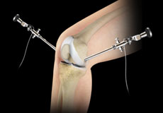
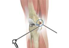
Knee Arthroscopy
Knee arthroscopy is a common surgical procedure performed using an arthroscope, a viewing instrument, to diagnose or treat a knee problem. It is a relatively safe procedure and you will usually be discharged from the hospital on the same day of surgery.
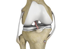
Multiligament Reconstruction of the Knee
Multiligament knee reconstruction is a surgical procedure to repair or replace two or more damaged ligaments of the knee joint. The surgery can be performed using minimally invasive techniques.
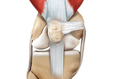
Posterolateral Corner (PLC) Reconstruction
A posterolateral corner is a complex arrangement of multiple ligaments, tendons, muscles and a joint capsule in your knee. It is located on the outside back corner of the knee.
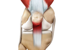
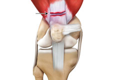
Quadriceps Tendon Rupture and Repair
The quadriceps tendon is a strong rope-like fibrous tissue located at the top of the patella or kneecap that connects the quadriceps muscles to the kneecap. It works together with the quadriceps muscles to allow us to straighten our leg.
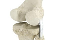
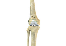
Knee Osteotomy
Knee osteotomy is a surgical procedure in which the upper part of shinbone (tibia) or lower part of thighbone (femur) is cut and realigned. It is usually performed in arthritic conditions affecting only one side of your knee.
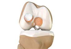
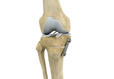
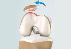
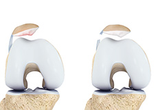
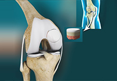
Cartilage Restoration of the Patellofemoral Joint
The articular or hyaline cartilage is the tissue lining the surface of the two bones in a joint. Cartilage helps the bones move smoothly against each other and can withstand the weight of your body during activities such as running and jumping.
Patellofemoral Realignment
Patellofemoral realignment is a surgical procedure performed to treat symptomatic patellofemoral instability that does not respond to nonsurgical treatment measures.

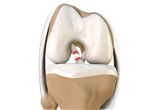
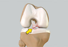
Primary ACL Repair
Traditionally, anterior cruciate ligament (ACL) tears of the knee are treated by reconstruction using a tendon or ligament autograft (tissue taken from your own body) or an allograft (tissue taken from a donor).
Lateral Lengthening
Lateral lengthening, also known as lateral retinacular lengthening or release, is a surgical procedure to release a tightened lateral retinaculum on the outer aspect of the knee.
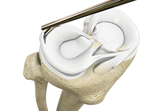
Meniscal Transplantation
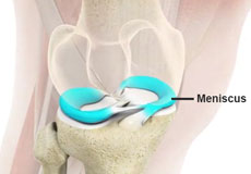
Meniscal transplantation is a surgical procedure to replace the damaged meniscus of the knee with healthy cartilage. The meniscus is a C-shaped cartilage ring that acts as a cushion between the shinbone and the thighbone.
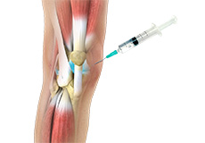
Viscosupplementation
Viscosupplementation refers to the injection of a hyaluronan preparation into the joint. Hyaluronan is a natural substance present in the joint fluid that assists in lubrication.
Distal Femoral Osteotomy
An osteotomy is a surgical procedure that involves cutting of bone. The distal femur is part of the femur (thighbone) just above the knee joint. Distal femoral osteotomy is performed to correct knee alignment which can lead to excessive loading and degeneration of one side of the knee joint.
Partial Meniscectom
Partial meniscectomy is a surgical procedure to remove the torn portion of the meniscus from the knee joint.
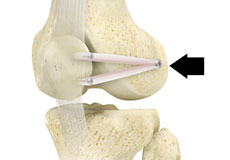
Failed Anterior Cruciate Ligament (ACL) Reconstruction
The knee joint is stabilized by four strong ligaments. The anterior cruciate ligament (ACL) passes diagonally in the middle of the knee, ensuring that the thigh and shin bone do not slide out of alignment during movement.
Medial Patellofemoral Ligament Reconstruction
Medial patellofemoral ligament reconstruction is a surgical procedure indicated for severe patellar instability.
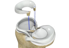
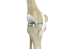
Arthroscopic Reconstruction of the Knee for Ligament Injuries
Arthroscopic knee ligament reconstruction is a surgical procedure to correct a torn knee ligament by replacing the ligament with a healthy tendon tissue using an arthroscope.
Knee Ligament Injuries
The knee is a hinge joint made up of two bones, the thighbone (femur) and shinbone (tibia). Ligaments are tough bands of tissue that connect one bone to another bone. The ligaments of the knee stabilize the knee joint.
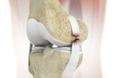
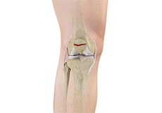
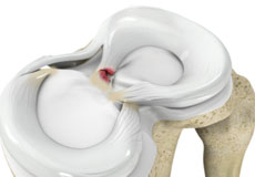
Meniscus Tears
A meniscal tear is a common knee injury in athletes, especially those involved in contact sports. A sudden bend or twist in your knee causes the meniscus to tear.
Patellar Instability
Any damage to the supporting ligaments may cause the patella to slip out of the groove either partially (subluxation) or completely (dislocation). This misalignment can damage the underlying soft structures such as muscles and ligaments that hold the kneecap in place.
Meniscal Injuries
Meniscal tears are one of the most common injuries to the knee joint. It can occur at any age but are more common in athletes involved in contact sports.
Knee Injury
Pain, swelling, and stiffness are the common symptoms of any damage or injury to the knee. If care is not taken during the initial phases of injury, it may lead to joint damage, which may end up destroying your knee.
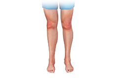
Knee Pain
Knee pain is a common condition affecting individuals of various age groups. It not only affects movement but also impacts your quality of life. An injury or disease of the knee joint or any structure surrounding the knee can result in knee pain.
Chondromalacia Patella
Chondromalacia patella is a common condition characterized by softening, weakening and damage of the cartilage. The condition is most often seen in young athletes and older adults who have arthritis of the knee.
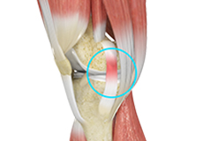
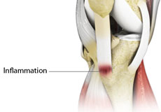
Jumper's Knee
Jumper’s knee, also known as patellar tendinitis, is inflammation of the patellar tendon that connects your kneecap (patella) to your shinbone. This tendon helps in the extension of the lower leg.
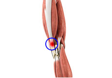
Iliotibial Band Syndrome
An iliotibial band is a tough group of fibers that runs from the iliac crest of the hip along the outside of the thigh, till the outer side of the shinbone, just below the knee joint.
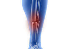
Fractures of the Tibia
The lower leg is made up of two long bones called the tibia and fibula that extend between the knee and ankle. The tibia or shinbone is the larger of the two bones. It bears most of the body’s weight and helps form the ankle joint and knee joint.
Osteochondritis Dissecans of the Knee
Osteochondritis dissecans is a joint condition in which a piece of cartilage, along with a thin layer of the bone separates from the end of the bone because of inadequate blood supply.
Pediatric ACL Tears
The anterior cruciate ligament (ACL) is a ligament that provides stability, reduces stress and prevents the knee from rotating or slipping out of position while jumping, running and landing.
Unstable Knee
Damage to any of these supportive structures causes instability of the knee joint. An unstable knee can be caused by the sudden twisting of the knee, tears of the meniscus, ligament
Knee Sprain
Knee sprain is a common injury that occurs from overstretching of the ligaments that support the knee joint. A knee sprain occurs when the knee ligaments are twisted or turned beyond its normal range, causing the ligaments to tear.
Multiligament Instability
The knee is a complex joint of the body that is vital for movement. The four major ligaments of the knee are anterior cruciate ligament (ACL), posterior cruciate ligament (PCL), medial collateral ligament (MCL) and lateral collateral ligament (LCL).
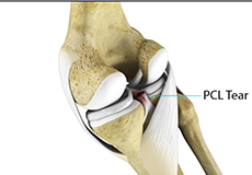
PCL Injuries
Posterior cruciate ligament (PCL), one of the four major ligaments of the knee, is situated at the back of the knee. It connects the thighbone (femur) to the shinbone (tibia). The PCL limits the backward motion of the shinbone.
MCL Sprains
Your MCL may get sprained or injured while twisting, bending or quickly changing direction. The medial collateral ligament (MCL), a band of tissue present on the inside of your knee joint, connects your thighbone and shinbone (bone of your lower leg).
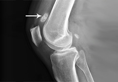
Patella Fracture
The kneecap or patella forms a part of the knee joint. It is present at the front of the knee, protecting the knee and providing attachment to various muscle groups of the thigh and leg.
Patellar Dislocation/Patellofemoral Dislocation
Patellar dislocation occurs when the patella moves out of the patellofemoral groove, (trochlea) onto the bony head of the femur. If the kneecap partially comes out of the groove, it is called subluxation
Quadriceps Tendon Rupture
The quadriceps can rupture after a fall, direct blow to the leg and when you land on your leg awkwardly from a jump. Quadriceps tendon rupture most commonly occurs in middle-aged people who participate in sports that involve jumping and running.
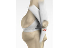
Patellar Tendon Rupture
The patellar tendon works together with the quadriceps muscle and the quadriceps tendon to allow your knee to straighten out. Patella tendon rupture is the rupture of the tendon that connects the patella (kneecap) to the top portion of the tibia
Patellofemoral Instability
Patellofemoral instability means that the patella (kneecap) moves out of its normal pattern of alignment. This malalignment can damage the underlying soft structures such as muscles and ligaments that hold the knee in place.
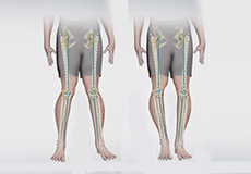
Knee Angular Deformities
Angular deformities of the knee are variations in the normal growth pattern during early childhood and are common during childhood. Physiologic angular deformities vary with age.
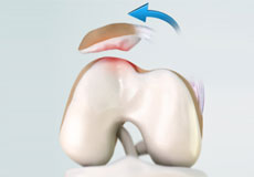
Articular Cartilage Injury
Articular or hyaline cartilage is the tissue lining the surface of the two bones in the knee joint. Cartilage helps the bones move smoothly against each other and can withstand the weight of the body during activities such as running and jumping.
Knee Sports Injuries
Trauma is any injury caused during physical activity, motor vehicle accidents, electric shock, or other activities. Sports trauma or sports injuries refer to injuries caused while playing indoor or outdoor sports and exercising.
Patellar Tendinitis
Patellar tendinitis, also known as "jumper's knee", is an inflammation of the patellar tendon that connects your kneecap (patella) to your shinbone. This tendon helps in extension of the lower leg.
Meniscus Root Tear
Meniscal root tears are characterized as soft tissue or bony root avulsion injuries or radial tears located within 1 cm of meniscus root attachment. They can be either a tear which disconnects the root area completely from the body of the meniscus
Osgood Schlatter Disease
Osgood-Schlatter disease refers to a condition in older children and teenagers caused by excessive stress to the patellar tendon (located below the kneecap). Participants in sports such as soccer, gymnastics, basketball, and distance running are at higher risk for this disease.
Multiligament Knee Injuries
Injury to more than one knee ligament is called a multiligament knee injury and may occur during sports or other physical activities.
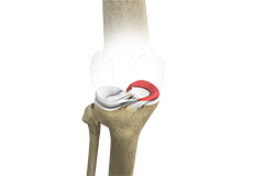
Patellofemoral Arthritis
Patellofemoral arthritis is an inflammatory condition characterized by loss of the smooth cartilage between the kneecap (patella) and the underlying femoral (thigh) bone in the knee joint.
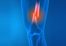
Stress Fracture of the Tibia
A stress fracture of the tibia or shinbone is a thin fracture, also called a hairline fracture that occurs in the tibia due to excess stress or overuse. The tibia is a weight-bearing bone in which stresses can accumulate from activities such as running and jumping.
Runner's Knee
Patellofemoral pain syndrome also called runner’s knee refers to pain under and around your kneecap. Patellofemoral pain is associated with a number of medical conditions such as anterior knee pain syndrome, patellofemoral malalignment, and chondromalacia patella.
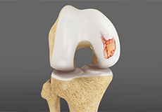
Chondral or Articular Cartilage Defects
The articular or hyaline cartilage is the tissue lining the surface of the two bones in the knee joint. Cartilage helps the bones move smoothly against each other and can withstand the weight of your body during activities such as running and jumping.
Recurrent Patella Dislocation
The patella (kneecap) is a small bone that shields your knee joint. It is present in front of your knee, on a groove called the trochlear groove that sits at the junction of the femur (thighbone) and tibia (shinbone).
Lateral Meniscus Syndrome
Lateral meniscus syndrome is characterized by an injury caused by the tearing of the cartilage tissue or a rare case of a congenital abnormality called a discoid meniscus, which results in knee pain.
Osteochondral Defect of the Knee
An osteochondral defect, also commonly known as osteochondritis dissecans, of the knee refers to a damage or injury to the smooth articular cartilage surrounding the knee joint and the bone underneath the cartilage.
Lateral Patellar Instability
Lateral patellar instability is defined as a lateral shift or displacement of the patella (kneecap) as a result of disruptive changes in the medial patellofemoral ligament (MPFL) and medial patellar retinaculum.
Medial Patellar Instability
Medial patellar instability is a disabling condition characterized by medial subluxation of the patella which occurs as a complication of lateral retinacular release surgery.
Patellar Tracking Disorder/Patellar Maltracking
Patellar tracking disorder, also known as patellar maltracking, is a condition in which the kneecap (patella) moves sideways from its groove when the leg is bent or straightened..
Loose Bodies in the Knee
Loose bodies are fragments of detached cartilage or bone inside the knee joint. These fragments may be free floating (unstable) or may be trapped (stable) within the joint.
Medial Meniscus Syndrome
Medial meniscal injuries are usually considered as either traumatic or degenerative. Whilst degenerate tears may present with a gradual history of increasing symptoms, traumatic injuries will usually occur as the knee is extended and rotated from a flexed position against resistance.
Knee Stress Fractures
Stress fractures of the patella or knee are very rare. Approximately two out of 10,000 athletes may experience a patella stress fracture.
Tibial Eminence Fractures
The tibia or shin bone is a major bone of the leg which connects the knee to the ankle. A fracture or break in the upper part of the tibia is known as a proximal tibial fracture and commonly occurs just below the knee joint.
Pediatric Tibial Tubercle Fractures
The tibia or shin bone is a major bone of the leg which connects the knee to the ankle. A fracture or break in the upper part of the tibia is known as a proximal tibial fracture and commonly occurs just below the knee joint.

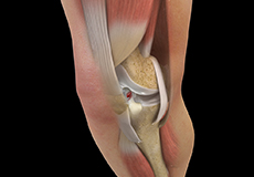
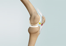
Knee Anatomy
The knee is a complex joint made up of different structures - bones, tendons, ligaments, and muscles. They all work together to maintain the knee’s normal function and provide stability to the knee during movement.
Having a well-functioning healthy knee is essential for our mobility and ability to participate in various activities. Understanding the anatomy of the knee enhances your ability to discuss and choose the right treatment procedure for knee problems with your doctor.
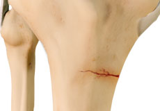
Bones of the Knee
The knee is a hinge joint made up of two bones, the thighbone (femur) and shinbone (tibia). There are two round knobs at the end of the femur called femoral condyles that articulate with the flat surface of the tibia called the tibial plateau. The tibial plateau on the inside of the leg is called the medial tibial plateau and on the outside of the leg, the lateral tibial plateau.
The two femoral condyles form a groove on the front (anterior) side of the knee called the patellofemoral groove. A small bone called the patella sits in this groove and forms the kneecap. It acts as a shield and protects the knee joint from direct trauma.
A fourth bone called the fibula is the other bone of the lower leg. This forms a small joint with the tibia. This joint has very little movement and is not considered a part of the main joint of the knee.
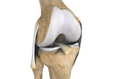
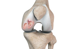
Articular Cartilage and Menisci of the Knee
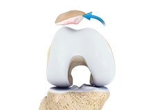
Movement of the bones causes friction between the articulating surfaces. To reduce this friction, all articulating surfaces involved in the movement are covered with a white, shiny, slippery layer called articular cartilage. The articulating surface of the femoral condyles, tibial plateaus and the back of the patella are covered with this cartilage. The cartilage provides a smooth surface that facilitates easy movement.
To further reduce friction between the articulating surfaces of the bones, the knee joint is lined by a synovial membrane that produces a thick clear fluid called synovial fluid. This fluid lubricates and nourishes the cartilage and bones inside the joint capsule.
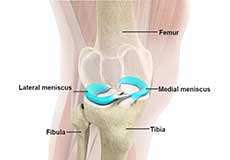
Within the knee joint, between the femur and tibia, are two C-shaped cartilaginous structures called menisci. Menisci function to provide stability to the knee by spreading the weight of the upper body across the whole surface of the tibial plateau. The menisci help in load-bearing i.e. it prevents the weight from concentrating onto a small area, which could damage the articular cartilage. The menisci also act as a cushion between the femur and tibia by absorbing the shock produced by activities such as walking, running and jumping.
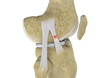
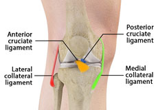
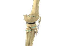
Ligaments of the Knee
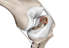
Ligaments are tough bands of tissue that connect one bone to another bone. The ligaments of the knee stabilize the knee joint. There are two important groups of ligaments that hold the bones of the knee joint together, collateral and cruciate ligaments.
Collateral ligaments are present on either side of the knee. They prevent the knee from moving too far during side to side motion. The collateral ligament on the inside is called the medial collateral ligament (MCL) and the collateral ligament on the outside is called the lateral collateral ligament (LCL).
Cruciate ligaments, present inside the knee joint, control the back-and-forth motion of the knee. The cruciate ligament in the front of the knee is called anterior cruciate ligament (ACL) and the cruciate ligament in the back of the knee is called posterior cruciate ligament (PCL).
Muscles of the Knee
There are two major muscles in the knee - the quadriceps and the hamstrings, which enable movement of the knee joint. The quadriceps muscles are located in front of the thigh. When the quadriceps muscles contract, the knee straightens. The hamstrings are located at the back of the thigh. When the hamstring muscles contract, the knee bends.
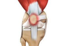
Tendons of the Knee
A tendon is a tissue that attaches a muscle to a bone. The quadriceps muscles of the knee meet just above the patella and attach to it through a tendon called the quadriceps tendon. The patella further attaches to the tibia through a tendon called the patella tendon. The quadriceps muscle, quadriceps tendon, and patellar tendon all work together to straighten the knee. Similarly, the hamstring muscles at the back of the leg are attached to the knee joint with the hamstring tendon.